
気流制限がある場合、呼気努力が生じる機序ー肺伸展受容体の機能からの考察
Blog-Dr城下の呼吸器雑記帳
「肺伸展受容体(Pulmonary stretch receptors)
これは気道平滑筋内に存在すると考えられ、肺の伸展に反応してインパルスを生ずる。肺が膨張している間はその活性は保たれ、適応による弱化は全くみられない。ここで発生したインパルスは、大有髄繊維を介して迷走神経内を伝導する。
これら受容体を刺激することによってえられる主な反射効果は、呼気時間の延長させることによって生じる呼吸数の減少である。これは、Hering-Breuer膨張反射として知られている。これはウサギを用いて、横隔膜の筋の一片から記録をうるようにして、他の呼吸筋からの影響を除外することによって明らかに証明することができる。古典的実験で、肺の膨張はそれ以上の吸気筋活性を抑制する傾向があることが知られていた。しかし、その逆の反応もみられ、すなわち、肺の縮小は吸気活動を惹起する傾向がある(縮小反射)。このように、これら反射は、自己調節機構ないしnegative feedbackを提供しうる。
このHering-Breuer反射は、一時は呼吸の数と深さを決定することにより換気における主要な役割を果たしていると考えられていた。これは、これらの伸展受容体からの情報を用いて延髄の“転換”機構を修飾することによって達成される。そしてまた、ほとんどの動物で、両側の迷走神経切断を行うと、緩徐で深い呼吸を生じる。しかし、より新しい研究によれば、この反射は成人では概して不活性であり、運動時のごとき一回換気量が1リットルを超える場合でもなければ作動しないことが示されている。覚醒しているヒトで局所麻酔により、一過性両側迷走神経ブロックを行なっても、呼吸数あるいは呼吸量に変化をきたさないことが知られている。しかしこれら反射は、新生児ではより重要な役割を果たしているだろうと考えられるいくつかの証拠がある。」1
「Hering-Breuerの反射
Heringたち(1865)は、肺には伸張受容器があって肺が拡張されるとそれが興奮して延髄にインパルスを送り、肺が縮小するとインパルス発射が止むことを発見した。彼らはこの求心性インパルスが吸息性ニューロンの持続的な活動を周期的に抑制することによって呼吸のリズムが形成されると考えた。
現在では肺を膨らませたとき吸息の抑制をきたす拡張反射inflation reflexと、逆に空気を抜いて肺を縮めたとき吸息の促進をきたす縮小反射deflation reflexとは別の反射と考えられている。しかし両者とも求心系は迷走神経を通るので、迷走神経を切ると吸息相も呼息相も延長する。それで両者をまとめてヘーリンク・ブロイエルの反射といっている。呼吸の切り換え反射(Shift-reflex)または呼吸運動の自動調節反射とも言われている。
この反射は動物では顕著で吸息の終期に吸息中枢抑制のため呼吸停止期がみられる。ヒトにもこの反射は存在するが、その作用は弱く、頸部迷走神経をブロックしても呼吸数はあまり変わらない。動物で迷走神経を左右とも切断すると呼吸のリズムは遅くなり、深くなる。しかし全く呼吸が停止してしまうことはない。」2
「Hering-Breuer reflexes
The basic rhythm of respiration may be modified so that the breathing rate, depth, or both are changed. The reason for the modification is to change the rate of ventilation in response to body needs. Afferent impulses to the respiratory center from several receptor sources have been identified. The most noteworthy of these among many of the animals are the Hering-Breuer reflexes. The receptors for these reflexes are located in the lung and particularly in the bronchi and bronchioles.
There are two components to the Hering-Breuer reflexes: (i) the inspiratory-inhibitory or inflation reflex and (ii) the inspiratory or deflation reflex. The nerve impulses generated by the receptors of the Hering-Breuer reflexes are transmitted by fibers in the vagus nerves to the respiratory center. The effect of inflation-receptor stimulation is to inhibit further inspiration (stimulation of neurons in the DRG) and to stimulate expiratory nerves in the VRG. The inspiratory or deflation reflex component is activated at some particular point of deflation. The deflation receptors might not be activated to bring about the next inspiration during eupnea, but they might be active when deflation is more complete. Deflation reflex receptor stimulation can be elicited in anesthetized dogs by manual compression of the thorax, which is followed immediately by inspiration. Practical use of this reflex is appropriate for respiratory depressed or unresponsive animals to promote more adequate ventilation in the former or to initiate ventilation in the latter. During exercise when tidal volume and frequency are increased, it would appear that the deflation reflex is more active in order to hasten the beginning of the next inspiration.
In addition to the receptors located in the lung, there are other peripherally located receptors that assist in modifying the basic rhythm. Stimulation of receptors in the skin is excitatory to the respiratory center, and deeper than usual inspiration may be noted. Perhaps their excitation to the inspiratory area is through the apneustic center, inasmuch as inspiratory gasps are occasionally seen. Advantage is taken of these receptors when stimulation of breathing is desired in newborn animals. Rubbing the skin with a rough cloth often starts the breathing cycles. An assist to ventilation needed during muscle activity is obtained from receptors located in tendons and joints. They will be stimulated when muscle contraction causes movement. It is also believed that when impulses are directed to skeletal muscles from the cerebral cortex, collateral impulses go to the brainstem and stimulate the respiratory center to increase alveolar ventilation. This mechanism might account for increases in ventilation that are not explainable by mere observation of changes in carbon dioxide, oxygen, and hydrogen ion concentration in the blood.」 3
考察
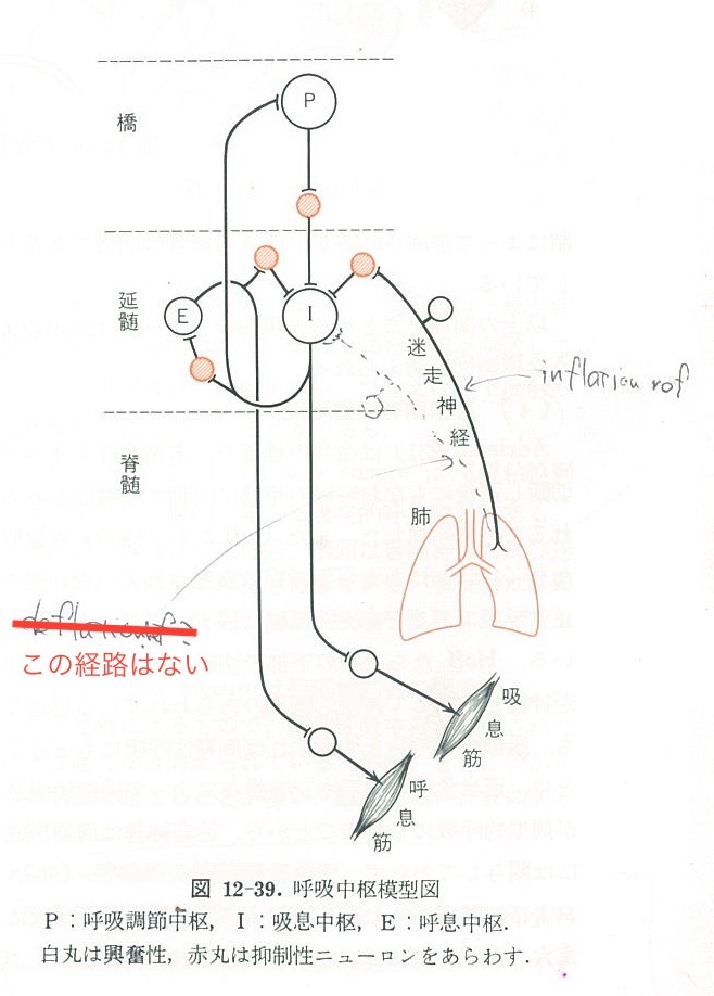
動物では、肺伸展受容体はヒトより強く作用し、膨張反射(気道圧が上がることに対する反応)には強く反応するようである。ここでいう「呼気時間の延長」が呼息筋系を刺激するかどうか明確には記述されていないが、膨張に対し強く吸息を抑制するのであれば、代わりに呼息中枢の活動を促進する可能性がある(以下、参考文献2 p343 図12-39を参照)。動物では(とくに犬でよく見られるが)、気流制限が生じている場合、同じ一回換気量でも末梢気道内抵抗が上昇し、それが肺伸展受容体を刺激し吸気抑制インパルスを強く生じ、結果として呼息筋運動を刺激する結果になると考えられる。
参考文献
1. West JB. 呼吸の生理. pp125-126, In: 笛木隆三,小林節雄訳, eds. 第2版 ed. 東京: 医学書院, 1989.
2. 真島英信. 生理学. pp342-343, 東京: 文光堂, 1986.
3. Reece WO. Regulation of Respiration. In: Reece WO, ed. Duke’s Physiology of Domestic Animals. 13th ed. Ames, IA: Wiley Blackwell, 2015;232-238.